When Mary Kananen teaches about cerebral circulation, there are times she gets a bit frustrated. It’s not because of the students but rather the limitations of how she can physically show them what she’s talking about.
“Cerebral circulation is a difficult concept for students to understand because it’s three-dimensional,” states Kananen, who is an associate teaching professor of biology. “It’s hard for students to visualize the way the arteries bend and wrap through the brain. I can tell them an artery turns and goes somewhere, but without being able to see it, it’s a challenging spatial relationship to figure out.”
Sure, there are models Kananen can use that show limited portions of the brain and some detail, and she has sometimes resorted to her own handmade polymer clay and pipe cleaner models—but of course, these are inadequate. And yes, there are complete models that would be perfect as teaching aids, but they aren’t readily available on the market, and, of course, they are incredibly expensive.
But at Penn State Altoona, a collaborative spirit and technology are transforming student learning experiences.
Here’s where Rebecca Strzelec and Penn State Altoona’s 3D printing lab come in.
Strzelec is a Distinguished Professor of visual arts at the college and often works with other departments on campus to create 3D models at The Center for Additive Manufacturing and Printing (CAMP).
The CAMP, established in 2017, is in the Doing Better Business 3D Printing Lab in the Eiche Library. It serves as a resource for students, faculty, and staff and supports 3D printing, 3D scanning, vinyl cutting, and large format inkjet printing for projects. The collaborative nature of the work benefits all disciplines across the campus.
Kananen approached Strzelec last summer asking if it were possible to print a 3D model of the cerebral arteries that weave throughout the brain. Kananen supplied Strzelec with a 3D model of the arteries, but they needed to be modified to suit Kananen’s needs. The data was correct in scale and location but needed significant refinement and repair in areas like connections and transitions. Further, some of the pieces would have been so small the model wouldn’t be able to hold itself and would break. Strzelec needed to increase the overall scale of the model so students could see what are actually hair-thin parts.
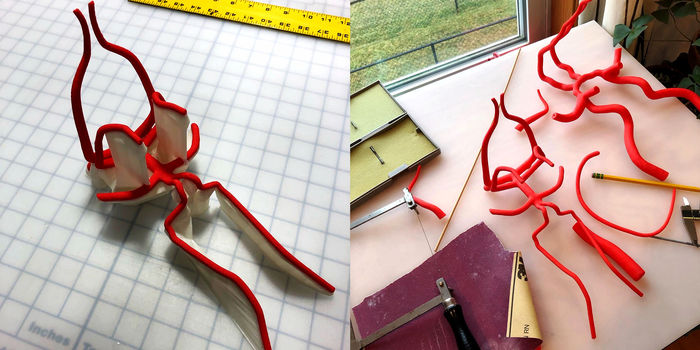
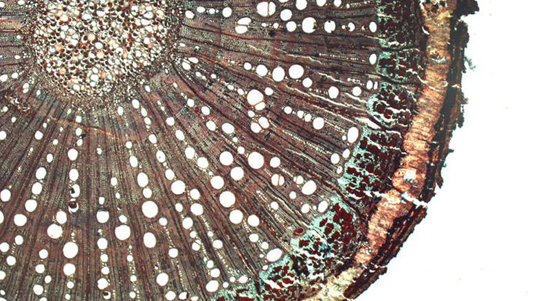
The cerebral artery model in various stages of completion
“It needed a lot of love and care,” says Strzelec of the project. “I started by evaluating the form of all pipe-like structures. It seemed odd that where limbs of the arteries joined it wasn’t more organic or smooth.” She consulted with Kananen who described that in the connection areas the arteries gradually flow into one another and that there are no hard edges or corners as shown in the model. That meant that Strzelec had to perform a sort of digital surgery. Each junction point had to be trimmed out and rebuilt so that they better resembled their true characteristics.
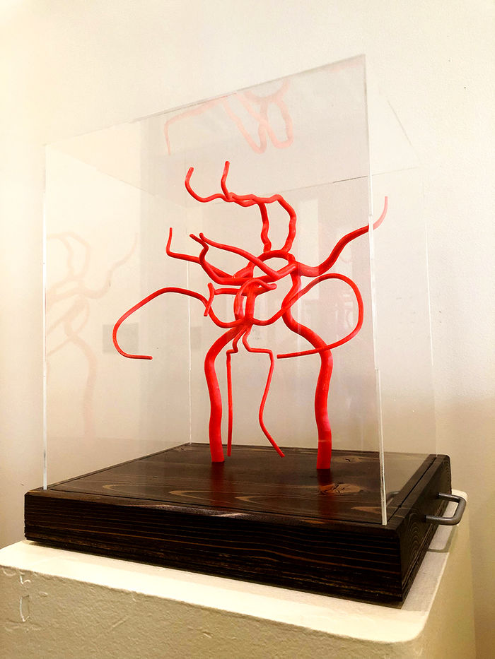
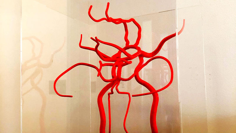
The completed model in its protective case
When Strzelec completed the modeling stage and then the 3D printing in a bright red ABS plastic, she turned to Penn State Altoona carpenter Tom Vogel to bring it all together. Vogel fabricated the wood base from plans pulled straight from the 3D modeling software. Working in this way allowed him to know exactly where to place the holes that slip into the base. According to Strzelec, “Tom’s craft is excellent, and at this point we’ve worked on enough projects together that he really understands the design and function of a build from just a five-minute meeting—he's terrific!” Strzelec then fabricated a clear acrylic case to protect the model from dust and damage.
The model will be used for students in anatomy, physiology, and pre-nursing courses as well as in some upper-level classes. “There is a team of us who will get to use it starting in the fall, and we are so excited,” says Kananen. “Rebecca’s background in this technology and her skill set is incredible. To me, what she’s done is beyond comprehension. This model will be an amazing resource for our students.”
Both professors have enjoyed working together and thought it was fun seeing things from each other’s academic perspective. Strzelec says she has worked on all sorts of projects with non-artists at Penn State Altoona, but this model may be the most intricate and wonderful yet.
“I love it, and I’m so glad I was able to do it. When it was about to be delivered, I was sad because I wanted to keep it. You surround yourself with things that bring you awe and joy, and when you figure out certain problems, you want evidence of that. But I can come visit it.” The model will live on the shelves in the labs of Holtzinger, tucked in safely near its new anatomical family.
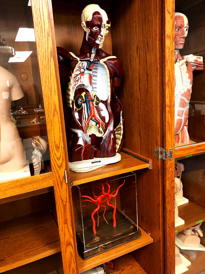
The completed model safely in its new home in Holtzinger
Up next for Strzelec is to create a training arena for a biology professor who is doing research on zebra fish. She loves the balance between working in her home art studio on her own research and creative practice and pivoting to help colleagues and students realize their projects via The CAMP.
“This kind of collaboration only happens here. I have never heard of a place where an art professor can do projects like this with a scientist this easily. There aren’t silos and walls here. The trust we have in each other’s abilities to create something is wonderful. The students get the biggest benefit from it, and that’s the overall mission. When given opportunities to work together, we pounce because we’re ready.”